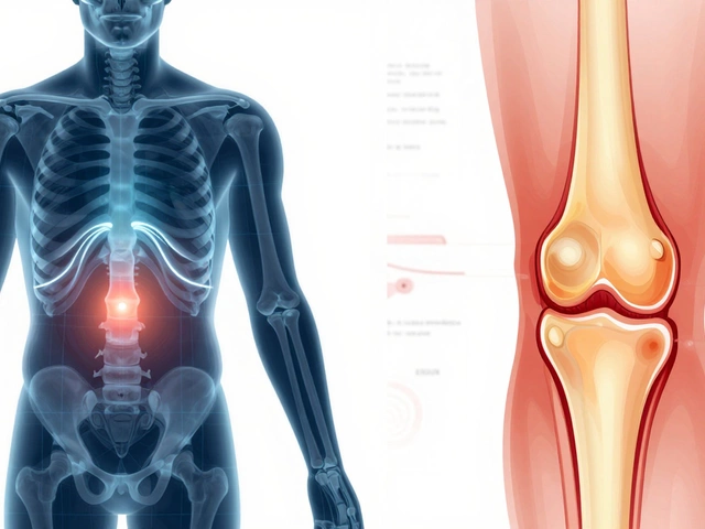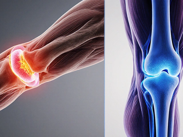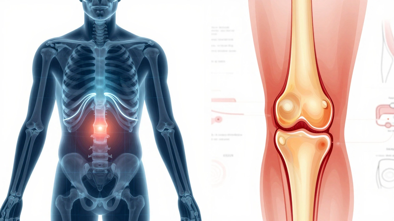
Orthopedic Imaging Decision Tool
Select the option that best describes your situation:
Recommended Approach
--
--
Select a symptom and click "Recommend Test" to see which imaging modality is typically best suited for your condition.
Waking up with a stiff knee or feeling a sharp twinge in your shoulder can be frustrating. You know something is wrong, but you don't know what. The first question that pops into your head is usually: "Which test should I get?" It’s a fair question because there isn’t one single test that works for every bone or joint problem. Choosing the right diagnostic tool depends entirely on what kind of injury or condition you suspect.
If you’ve ever been told to "get an X-ray" without much explanation, you might feel like it’s just a standard procedure. But in orthopedics, imaging is precise. An X-ray is a quick, low-cost imaging technique that uses radiation to visualize dense structures like bones is great for fractures. It falls short when it comes to soft tissues like ligaments or cartilage. On the other hand, an MRI (Magnetic Resonance Imaging) provides detailed images of soft tissues, including muscles, tendons, ligaments, and cartilage, using magnetic fields and radio waves is the gold standard for those areas. Understanding the difference between these tools helps you advocate for yourself during your doctor’s visit.
The First Line of Defense: X-Rays
When you walk into an orthopedic clinic with acute pain after a fall or accident, the doctor will almost always start with an X-ray. Why? Because speed and clarity matter most in emergencies. X-rays are excellent at showing broken bones, dislocations, and signs of arthritis. They take seconds to perform and cost significantly less than advanced scans.
However, X-rays have blind spots. They cannot show you a torn ACL (anterior cruciate ligament), a meniscus tear in the knee, or inflammation in the joint lining. If your X-ray comes back "normal" but you’re still in pain, don’t assume nothing is wrong. It just means the problem lies in the soft tissue, which requires a different type of imaging.
- Best for: Fractures, bone tumors, severe osteoarthritis, spinal alignment issues.
- Limitations: Poor visibility of soft tissues; involves ionizing radiation.
- Cost & Time: Low cost; results available within minutes.
Seeing the Soft Tissue: MRI Scans
If your pain is chronic, or if you suspect a sports injury involving ligaments or tendons, an MRI is likely the next step. Unlike X-rays, MRIs do not use radiation. Instead, they use strong magnetic fields to align hydrogen atoms in your body, creating incredibly detailed cross-sectional images. This makes them indispensable for diagnosing complex joint issues.
For example, if you’re a runner with persistent knee pain, an MRI can reveal a subtle tear in the meniscus that an X-ray would miss entirely. It’s also the preferred method for evaluating nerve compression in the spine, such as a herniated disc pressing on a nerve root. While MRIs are more expensive and take longer (usually 30-60 minutes), the detail they provide often changes the treatment plan from physical therapy to surgery, or vice versa.
- Best for: Ligament tears, tendon injuries, cartilage damage, spinal disc herniation, brain and spinal cord abnormalities.
- Limitations: Higher cost; claustrophobia can be an issue; metal implants may interfere with the scan.
- Cost & Time: Moderate to high cost; takes 30-60 minutes.
Complex Structures and Surgery Planning: CT Scans
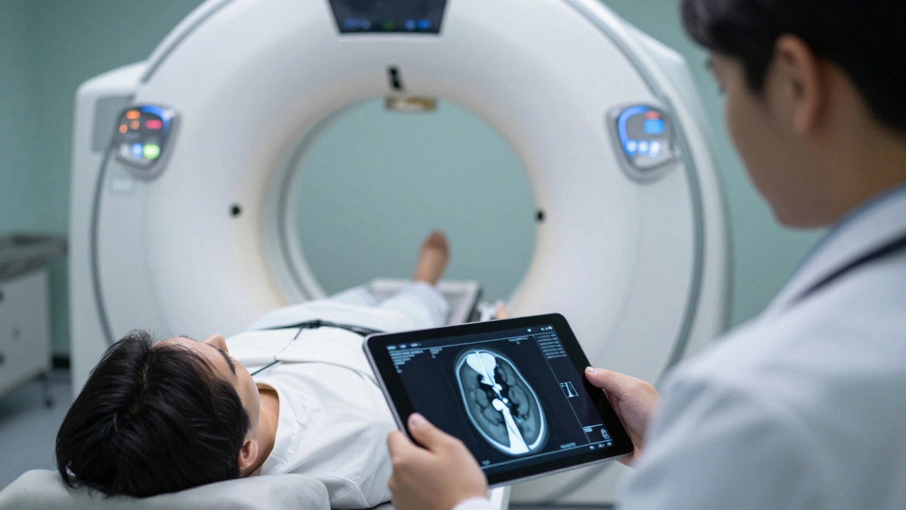
Sometimes, an X-ray is too vague, and an MRI is not necessary for bone structure alone. That’s where a CT Scan (Computed Tomography) comes in. Think of a CT scan as a series of X-rays taken from different angles and combined by a computer to create detailed 3D images. This technology is particularly useful for complex fractures, such as those in the pelvis, spine, or wrist, where surgeons need to see exactly how the bone fragments align before operating.
CT scans are also faster than MRIs, making them ideal for patients who cannot lie still for long periods. However, because they use higher doses of radiation than standard X-rays, doctors reserve them for cases where the additional detail is critical for surgical planning or when metal implants prevent an MRI.
- Best for: Complex fractures, pre-surgical planning, detecting small bone lesions, patients with metal implants who cannot undergo MRI.
- Limitations: Higher radiation exposure than X-rays; less effective for soft tissue than MRI.
- Cost & Time: Moderate cost; takes 10-30 minutes.
Real-Time Movement: Ultrasound
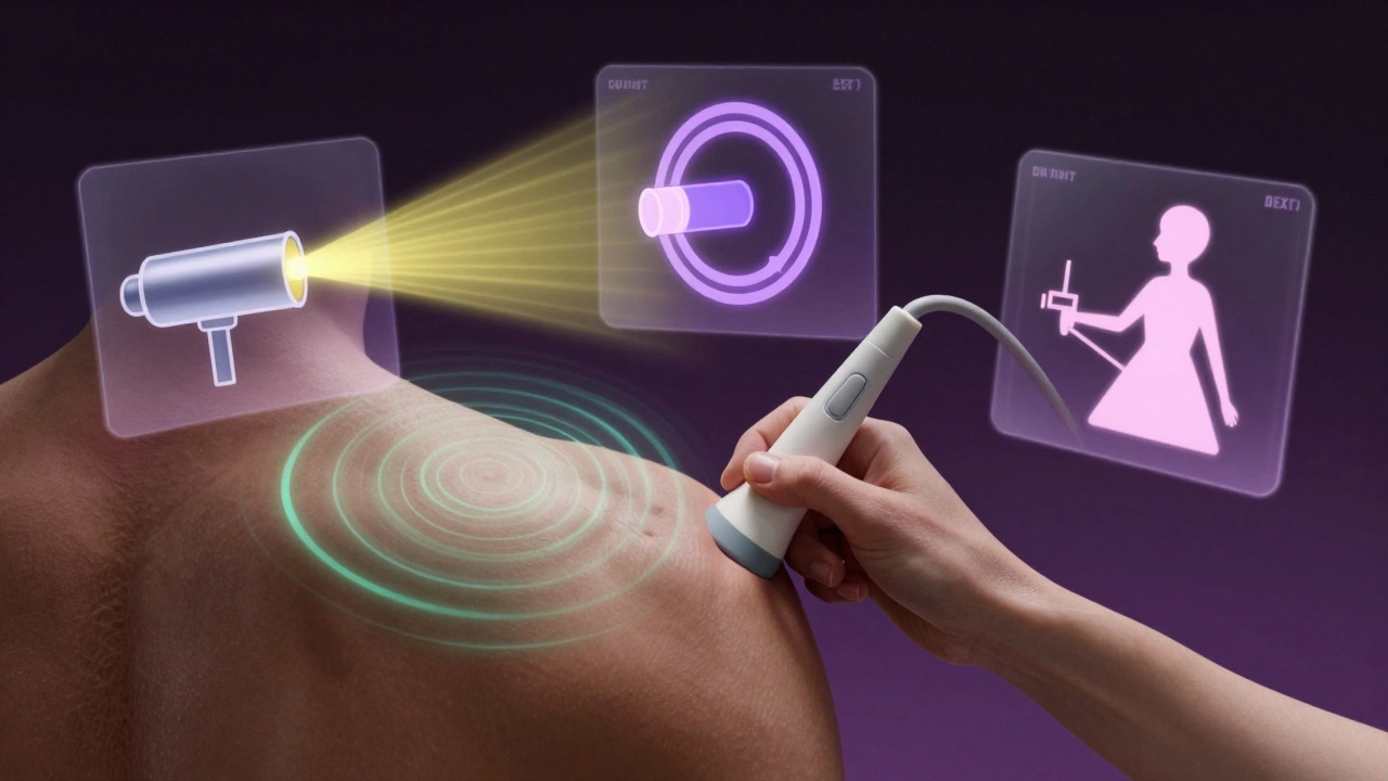
Ultrasound is often overlooked in orthopedics, but it has a unique advantage: dynamic imaging. While X-rays, MRIs, and CTs show static pictures, ultrasound allows the doctor to see the joint moving in real-time. This is incredibly useful for assessing shoulder impingement, rotator cuff tears, or tendonitis in the elbow or ankle.
Because it uses sound waves instead of radiation, ultrasound is safe for repeated use and even for pregnant women. It’s also portable, meaning specialists can bring the machine directly to your consultation room. If you have superficial joint pain-like in the shoulder, elbow, or knee-ultrasound can provide immediate answers without the wait time of an MRI scheduling backlog.
- Best for: Superficial tendon and ligament injuries, guiding injections, assessing fluid accumulation (bursitis), pediatric patients.
- Limitations: Cannot penetrate deep joints (like the hip) or bone; operator-dependent quality.
- Cost & Time: Low to moderate cost; immediate results.
Bone Strength Assessment: DEXA Scans
If your concern is not about an acute injury but rather long-term bone health, especially if you are over 50 or have risk factors for osteoporosis, a DEXA Scan (Dual-Energy X-ray Absorptiometry) is the best test. This specialized X-ray measures bone mineral density (BMD). It doesn’t look for breaks or tears; it looks for weakness.
A DEXA scan is crucial for preventing future fractures. By identifying osteopenia (low bone mass) or osteoporosis early, doctors can prescribe calcium, vitamin D, or medication to strengthen your bones before a minor fall leads to a major break. It’s a quick, non-invasive procedure that typically focuses on the hip and spine, the most common sites for osteoporotic fractures.
- Best for: Diagnosing osteoporosis, monitoring bone loss over time, assessing fracture risk.
- Limitations: Does not detect fractures or soft tissue injuries; limited to specific skeletal sites.
- Cost & Time: Low cost; takes 10-20 minutes.
| Test Type | Primary Use | Radiation Exposure | Average Cost (India) | Time Required |
|---|---|---|---|---|
| X-Ray | Fractures, Arthritis | Low | ₹500 - ₹1,500 | 5-10 mins |
| MRI | Ligaments, Cartilage, Spine | None | ₹3,000 - ₹8,000 | 30-60 mins |
| CT Scan | Complex Fractures, Surgery Planning | High | ₹2,000 - ₹5,000 | 10-30 mins |
| Ultrasound | Tendons, Real-time Movement | None | ₹1,000 - ₹3,000 | 15-30 mins |
| DEXA Scan | Bone Density, Osteoporosis | Very Low | ₹1,500 - ₹3,000 | 10-20 mins |
How to Choose the Right Test?
You shouldn’t have to guess which test you need. Your orthopedic specialist will decide based on your symptoms, medical history, and physical examination. Here is a simple decision tree to understand their logic:
- Did you hear a pop or snap? This often indicates a ligament or tendon tear. An MRI or Ultrasound is likely needed.
- Did you fall or hit the area? Start with an X-ray to rule out fractures. If the X-ray is clear but pain persists, move to MRI.
- Is the pain gradual and achy? This could be arthritis or degenerative changes. An X-ray is usually sufficient to assess joint space narrowing.
- Are you over 50 with back or hip pain? Consider a DEXA scan to check for osteoporosis, especially if you have a family history.
- Do you have metal implants? Inform your doctor immediately. You may need a CT scan instead of an MRI.
Remember, no test is "best" in isolation. The best test is the one that answers your specific clinical question. Over-testing can lead to unnecessary anxiety and costs, while under-testing can delay proper treatment. Trust your doctor’s judgment, but don’t hesitate to ask why a particular test is recommended.
Is an MRI better than an X-ray for joint pain?
Not necessarily. X-rays are better for viewing bone structure, fractures, and arthritis. MRIs are superior for visualizing soft tissues like ligaments, tendons, and cartilage. For general joint pain, doctors often start with an X-ray because it’s cheaper and faster. If the X-ray is normal but pain continues, an MRI is then ordered to check for soft tissue damage.
Can an ultrasound replace an MRI for knee pain?
In some cases, yes. Ultrasound is excellent for superficial structures like the patellar tendon or bursae. However, it cannot see deep inside the knee joint to evaluate the meniscus or cruciate ligaments effectively. For comprehensive knee assessment, MRI remains the gold standard.
How often can I have a DEXA scan?
DEXA scans involve very low radiation, so they are safe for regular monitoring. Doctors typically recommend repeating the scan every 1-2 years if you are being treated for osteoporosis, or every 5+ years if you are at average risk. Frequent scanning is unnecessary unless your treatment plan changes significantly.
Why did my doctor order a CT scan instead of an MRI?
This usually happens for two reasons: either you have a metal implant (like a pacemaker or certain joint replacements) that makes MRI unsafe, or the doctor needs a detailed 3D view of complex bone fractures for surgical planning. CT scans are also faster, which helps patients who struggle to stay still for long periods.
Do I need contrast dye for these tests?
It depends. X-rays and standard CT scans rarely need contrast. MRIs sometimes use gadolinium-based contrast agents to highlight inflammation, tumors, or infections. Ultrasounds may use saline solution for hydrodissection during guided injections. Always inform your doctor about kidney function or allergies before receiving any contrast material.